
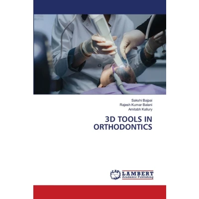
3D TOOLS IN ORTHODONTICS
Uitgelicht

|
47,99 |
Naar shop
|

|
48,60 |
Naar shop
|

|
48,60 |
Naar shop
|
| VERGELIJK ALLE AANBIEDERS (3) | ||
 Beschrijving
Bol
Three-dimensional (3D) imaging tools have brought significant advancements in orthodontic diagnosis, treatment planning, and evaluation of treatment outcomes. Traditionally, orthodontic records were based on two-dimensional (2D) methods such as cephalometric radiographs, panoramic radiographs, clinical photographs, and plaster models. Although these methods have been widely used, they possess inherent limitations, including image distortion, projection errors, inconsistent landmark identification, and difficulty in accurately evaluating complex craniofacial structures. The conversion of three-dimensional anatomical structures into two-dimensional images frequently results in the loss of important diagnostic information and may obscure asymmetries.The introduction of 3D imaging technologies has enabled clinicians to visualize craniofacial structures in their true anatomical dimensions, thereby improving diagnostic accuracy and treatment planning.


 Beschrijving
(1)
Beschrijving
(1)
Three-dimensional (3D) imaging tools have brought significant advancements in orthodontic diagnosis, treatment planning, and evaluation of treatment outcomes. Traditionally, orthodontic records were based on two-dimensional (2D) methods such as cephalometric radiographs, panoramic radiographs, clinical photographs, and plaster models. Although these methods have been widely used, they possess inherent limitations, including image distortion, projection errors, inconsistent landmark identification, and difficulty in accurately evaluating complex craniofacial structures. The conversion of three-dimensional anatomical structures into two-dimensional images frequently results in the loss of important diagnostic information and may obscure asymmetries.The introduction of 3D imaging technologies has enabled clinicians to visualize craniofacial structures in their true anatomical dimensions, thereby improving diagnostic accuracy and treatment planning.
 Productspecificaties
 Prijshistorie
* Prijshistorie bevat geen data van Amazon, Amazon Marketplace.
Prijzen voor het laatst bijgewerkt op:
 Gerelateerde resultaten


