
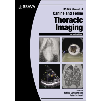
BSAVA Manual of Canine and Feline Thoracic Imaging
Uitgelicht

|
109,00 |
Naar shop
|
| VERGELIJK ALLE AANBIEDERS (1) | ||
 Beschrijving
Bol
This new edition provides a comprehensive textbook on diagnostic imaging of the canine and feline thorax. The Manual includes dedicated sections on the principles of thoracic imaging interpretation and individual body systems, clearly structured for information retrieval. High-quality images and illustrations demonstrate normal radiographic appearance and abnormalities associated with disease. The second edition adds new scientific knowledge, mainly gained in CT and MRI including knowledge that can be applied to radiographic interpretation, still the most widely used imaging modality for this body system.


Vergelijk aanbieders (1)

 Beschrijving
(1)
Beschrijving
(1)
This new edition provides a comprehensive textbook on diagnostic imaging of the canine and feline thorax. The Manual includes dedicated sections on the principles of thoracic imaging interpretation and individual body systems, clearly structured for information retrieval. High-quality images and illustrations demonstrate normal radiographic appearance and abnormalities associated with disease. The second edition adds new scientific knowledge, mainly gained in CT and MRI including knowledge that can be applied to radiographic interpretation, still the most widely used imaging modality for this body system.
 Productspecificaties
| EAN |
|
|---|---|
| Maat |
|
 Prijshistorie
Prijzen voor het laatst bijgewerkt op:
 Gerelateerde resultaten


