
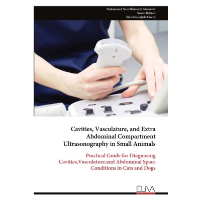
Cavities, Vasculature, and Extra Abdominal Compartment Ultrasonography in Small Animals
Uitgelicht

|
59,99 |
Naar shop
|

|
74,20 |
Naar shop
|
| VERGELIJK ALLE AANBIEDERS (2) | ||
 Beschrijving
Bol
Cavities, Vasculature, and Extra-Abdominal Compartment Ultrasonography in Small Animals is a comprehensive, practical guide designed to expand the diagnostic capabilities of veterinary professionals working with dogs and cats. Moving beyond the commonly imaged abdominal organs, this resource covers a broad range of anatomical regions, including the pleural, retroperitoneal, pericardial, pelvic, and umbilical cavities, along with essential vascular structures such as the abdominal aorta, caudal vena cava, portal vein, and the hepatic, renal, and mesenteric vessels.Each chapter follows a clear, structured format-beginning with a review of normal anatomy and ultrasonographic appearances, then progressing to common abnormalities, key differential diagnoses, and recommendations for additional diagnostics or therapeutic options.The text also explores less frequently discussed but clinically important areas, such as the perineal and inguinal regions, mammary and submammary tissues, and external genital structures including the scrotum, vulva, and prepuce. Special attention is given to variations in ultrasonographic appearance due to breed, age, and physiological state, ensuring clinical applicability across diverse patient populations.Designed for both new and experienced veterinarians, radiologists, and veterinary technicians, this guide bridges the gap between theoretical knowledge and clinical practice. Standardized scanning protocols, probe selection recommendations, and guidance on recognizing and managing common imaging artifacts enable practitioners to acquire high-quality images and extract meaningful diagnostic insights.


 Beschrijving
(1)
Beschrijving
(1)
Cavities, Vasculature, and Extra-Abdominal Compartment Ultrasonography in Small Animals is a comprehensive, practical guide designed to expand the diagnostic capabilities of veterinary professionals working with dogs and cats. Moving beyond the commonly imaged abdominal organs, this resource covers a broad range of anatomical regions, including the pleural, retroperitoneal, pericardial, pelvic, and umbilical cavities, along with essential vascular structures such as the abdominal aorta, caudal vena cava, portal vein, and the hepatic, renal, and mesenteric vessels.Each chapter follows a clear, structured format-beginning with a review of normal anatomy and ultrasonographic appearances, then progressing to common abnormalities, key differential diagnoses, and recommendations for additional diagnostics or therapeutic options.The text also explores less frequently discussed but clinically important areas, such as the perineal and inguinal regions, mammary and submammary tissues, and external genital structures including the scrotum, vulva, and prepuce. Special attention is given to variations in ultrasonographic appearance due to breed, age, and physiological state, ensuring clinical applicability across diverse patient populations.Designed for both new and experienced veterinarians, radiologists, and veterinary technicians, this guide bridges the gap between theoretical knowledge and clinical practice. Standardized scanning protocols, probe selection recommendations, and guidance on recognizing and managing common imaging artifacts enable practitioners to acquire high-quality images and extract meaningful diagnostic insights.
 Productspecificaties
 Prijshistorie
* Prijshistorie bevat geen data van Amazon.
Prijzen voor het laatst bijgewerkt op:
 Gerelateerde resultaten


