


€ 149,00 Gratis verzending

Uitgelicht
|
|
149,00 |
Naar shop
|

|
159,98 |
Naar shop
|

|
159,98 |
Naar shop
|
| VERGELIJK ALLE AANBIEDERS (3) | ||
 Beschrijving
Vergelijk aanbieders (3)
€ 149,00 Gratis verzending
€ 159,98 Gratis verzending
€ 159,98 Gratis verzending
Winkels met andere maten
Beschrijving
(1)
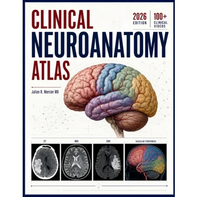
Students and residents often recognize neurologic syndromes only after seeing the scan. That delay weakens examination-based reasoning and makes lesion localization feel harder than it is.Clinical Neuroanatomy Atlas is the 2026 edition built to reverse that habit through cortical mapping, pathway tracing, lesion localization, diagnostic reasoning, and neuroradiological correlation that stays tied to real bedside findings.This atlas pairs structured clinical chapters with labeled illustrations, imaging-oriented explanations, and a QR-linked companion course with 100+ videos for reinforcement during clerkships, call nights, and board review.- 25 chapters covering cortex, deep nuclei, white matter tracts, cranial nerves, brainstem, spinal cord, and vascular territories- Lesion-first teaching that connects symptoms, signs, and cross-sectional imaging- Clinical images and high-yield tables designed for medical students and residents- Case-based diagnostic reasoning that shows how experts revise localization when new data arrivesUse this atlas to localize faster, read neuroradiology more intelligently, and connect every neuroanatomic structure to a clinical pattern that matters.
 Prijshistorie
 * Prijshistorie bevat geen data van Amazon, Amazon Marketplace.
Prijzen voor het laatst bijgewerkt op:
 Gerelateerde resultaten
 Productspecificaties


