
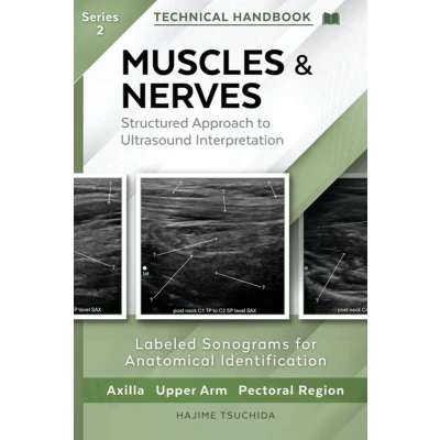
MUSCLES & NERVES - Technical Ultrasound Handbook of the Axilla, Upper Arm and Pectoral Region (Series 2): Structured Approach to Interpretation
Uitgelicht

|
60,99 |
Naar shop
|

|
78,98 |
Naar shop
|

|
78,98 |
Naar shop
|
| VERGELIJK ALLE AANBIEDERS (3) | ||
 Beschrijving
Bol
* Master Musculoskeletal Ultrasound Anatomy: Delve into detailed ultrasound anatomy of muscles and peripheral nerves.* Practical Scan Techniques: Gain technical explanations on how to acquire specific ultrasound structures.* Step-by-Step Guidance: Follow along with clear, step-by-step tracing of anatomical structures.* Labeled Ultrasound Images: Easily identify structures with precisely labeled ultrasound images.* Focused Visual Learning: Benefit from a unique "one page - one image" format for clear, concise understanding.* Your Essential Practical Handbook: Designed exclusively for hands-on, technical application.A practical visual atlas for clinicians performing musculoskeletal ultrasound and nerve tracing.A Visual, Step-by-Step Ultrasound Atlas for the Axilla, Upper Arm and Pectoral Region.This series is based on a dynamic scan sequence in which each image represents a continuous stage of probe movement rather than an isolated static view. By observing the sequential changes in anatomical structures during probe translation, readers can appreciate spatial relationships in a way that closely simulates real-time scanning.Whether you are performing MSK ultrasound, guiding nerve blocks, planning rehabilitation, or improving diagnostic understanding, this handbook supports clinical practice through clear anatomical reference-not as a substitute for medical diagnosis or treatment.WHAT YOU WILL LEARN: - How to confidently identify peripheral nerves and muscle layers on ultrasound- How to follow each structure in continuity, using step-by-step tracing guidance- How to differentiate muscles, fascial compartments, and branching nerve patterns- How to apply practical probe positioning techniques to achieve optimized imagesWHY THIS ATLAS IS UNIQUE: - "One Page - One Image" layout allows rapid comprehension during live scanning- Each ultrasound image is paired with a precise anatomical tracing for instant orientation- Structured for real-world scanning workflows, not textbook theoryWHO THIS BOOK IS FOR: - Sonographers and MSK imaging clinicians- Physical and Occupational Therapists- Orthopedics providers- Pain medicine and regional anesthesia clinicians- Students developing foundational sonoanatomy skillsA practical, visual, and clinically focused guide to mastering ultrasound anatomy of the Axilla, Upper Arm and Pectoral Region.


 Beschrijving
(1)
Beschrijving
(1)
* Master Musculoskeletal Ultrasound Anatomy: Delve into detailed ultrasound anatomy of muscles and peripheral nerves.* Practical Scan Techniques: Gain technical explanations on how to acquire specific ultrasound structures.* Step-by-Step Guidance: Follow along with clear, step-by-step tracing of anatomical structures.* Labeled Ultrasound Images: Easily identify structures with precisely labeled ultrasound images.* Focused Visual Learning: Benefit from a unique "one page - one image" format for clear, concise understanding.* Your Essential Practical Handbook: Designed exclusively for hands-on, technical application.A practical visual atlas for clinicians performing musculoskeletal ultrasound and nerve tracing.A Visual, Step-by-Step Ultrasound Atlas for the Axilla, Upper Arm and Pectoral Region.This series is based on a dynamic scan sequence in which each image represents a continuous stage of probe movement rather than an isolated static view. By observing the sequential changes in anatomical structures during probe translation, readers can appreciate spatial relationships in a way that closely simulates real-time scanning.Whether you are performing MSK ultrasound, guiding nerve blocks, planning rehabilitation, or improving diagnostic understanding, this handbook supports clinical practice through clear anatomical reference-not as a substitute for medical diagnosis or treatment.WHAT YOU WILL LEARN: - How to confidently identify peripheral nerves and muscle layers on ultrasound- How to follow each structure in continuity, using step-by-step tracing guidance- How to differentiate muscles, fascial compartments, and branching nerve patterns- How to apply practical probe positioning techniques to achieve optimized imagesWHY THIS ATLAS IS UNIQUE: - "One Page - One Image" layout allows rapid comprehension during live scanning- Each ultrasound image is paired with a precise anatomical tracing for instant orientation- Structured for real-world scanning workflows, not textbook theoryWHO THIS BOOK IS FOR: - Sonographers and MSK imaging clinicians- Physical and Occupational Therapists- Orthopedics providers- Pain medicine and regional anesthesia clinicians- Students developing foundational sonoanatomy skillsA practical, visual, and clinically focused guide to mastering ultrasound anatomy of the Axilla, Upper Arm and Pectoral Region.
 Productspecificaties
 Prijshistorie
* Prijshistorie bevat geen data van Amazon, Amazon Marketplace.
Prijzen voor het laatst bijgewerkt op:
 Gerelateerde resultaten


