

€ 77,75 Gratis verzending

Uitgelicht
|
|
77,75 |
Naar shop
|

|
83,99 |
Naar shop
|
| VERGELIJK ALLE AANBIEDERS (2) | ||
 Beschrijving
Vergelijk aanbieders (2)
€ 77,75 Gratis verzending
€ 83,99 Gratis verzending
Winkels met andere maten
Beschrijving
(1)
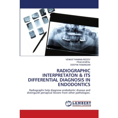
Radiographic interpretation and differential diagnosis are vital in endodontics for accurate diagnosis, treatment planning, and evaluation of outcomes. The advancement of dental radiology from conventional film to digital systems has improved image quality, reduced radiation dose, and increased diagnostic efficiency. Proper principles of radiographic selection and interpretation help in choosing the most appropriate imaging method while minimizing patient exposure.Radiographs are routinely used to assess root canal anatomy, detect periapical pathology, determine working length, identify additional canals, and evaluate root resorption. Image quality is affected by exposure factors, angulation, receptor placement, processing or sensor errors, and patient cooperation. Common techniques such as paralleling and bisecting angle methods are used before, during, and after treatment to assess canal preparation, obturation quality, procedural errors, and periapical healing. Recent advancements such as digital radiography and CBCT have enhanced diagnostic accuracy and improved endodontic decision making.
 Prijshistorie
 * Prijshistorie bevat geen data van Amazon.
Prijzen voor het laatst bijgewerkt op:
 Gerelateerde resultaten
 Productspecificaties


