


€ 87,90 Gratis verzending

Uitgelicht
|
|
87,90 |
Naar shop
|

|
113,91 |
Naar shop
|

|
113,91 |
Naar shop
|
| VERGELIJK ALLE AANBIEDERS (3) | ||
 Beschrijving
Vergelijk aanbieders (3)
€ 87,90 Gratis verzending
€ 113,91 € 4,49 verzendkosten Totaal € 118,40
€ 113,91 € 6,49 verzendkosten Totaal € 120,40
Winkels met andere maten
Beschrijving
(1)
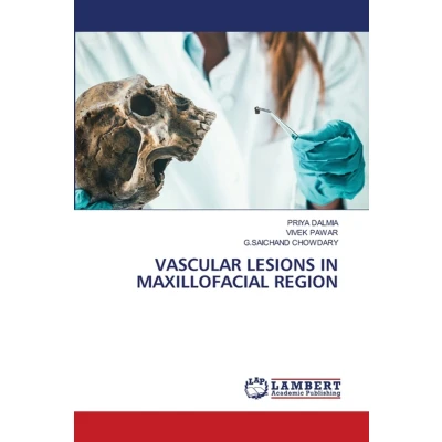
Vascular lesions of maxillofacial region include vascular tumors (hemangiomas) and vascular malformations, as classified by the ISSVA system. Hemangiomas are benign endothelial tumors that appear after birth, proliferate rapidly, and usually regress spontaneously, whereas vascular malformations are congenital structural abnormalities that persist throughout life and enlarge with growth, trauma, or hormonal changes. They commonly involve the head and neck and may cause pain, bleeding, facial deformity, functional impairment, and, in severe cases, life-threatening hemorrhage. Accurate diagnosis relies on clinical examination, Doppler ultrasonography, MRI, CT, angiography, and histopathology, with MRI serving as the gold standard for lesion evaluation. Recent advances in molecular genetics and imaging have improved diagnostic accuracy and treatment planning. Management is individualized according to lesion type and flow characteristics, using observation, medical therapy, sclerotherapy, laser therapy, embolization, and surgical excision. A multidisciplinary approach is essential for achieving optimal functional and aesthetic outcomes while minimizing complications.
 Prijshistorie
 * Prijshistorie bevat geen data van Amazon Marketplace, Amazon.
Prijzen voor het laatst bijgewerkt op:
 Gerelateerde resultaten
 Productspecificaties


