


€ 175,00 Gratis verzending

Uitgelicht
|
|
175,00 |
Naar shop
|
| VERGELIJK ALLE AANBIEDERS (1) | ||
 Beschrijving
Vergelijk aanbieders (1)
€ 175,00 Gratis verzending
Winkels met andere maten
Beschrijving
(1)
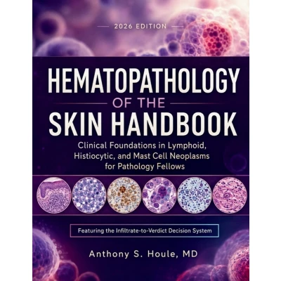
Read the Infiltrate, Reach the Verdict, Defend It at Sign-OutThis reference connects the normal immune architecture of skin to the decisions that separate a reactive lymphoid infiltrate from early lymphoma, moving from density and zonation at the microscope through staged immunohistochemistry and molecular clonality into the full range of cutaneous T-cell and B-cell lymphomas, histiocytic and dendritic cell disorders, and mast cell disease. Built for the dermatopathology fellow, the general pathologist without subspecialty backup, and the hematopathologist covering a skin consult, it treats every entity as a placement problem: where a specific infiltrate actually sits, and what one additional stain changes about that placement.A five-part diagnostic callout repeated across every major entity, consolidated into a forty-entry cross-referenced Atlas, turns pattern recognition under uncertainty into a repeatable, defensible sequence.Inside, You Will Learn How To- Separate reactive from neoplastic fast - density, zonation, and epidermotropism-to-spongiosis reasoning that catches early mycosis fungoides before it's missed.- Stage a lymphoid infiltrate without guessing - staged immunohistochemistry and clonality testing, including the pseudoclonality safeguard against false-positive lymphoma calls.- Place a CD30-positive lesion correctly - lymphomatoid papulosis versus anaplastic large cell lymphoma, resolved by clinical course and staging, not morphology alone.- Read the BCL2 reversal - why the same marker confirms malignancy in one B-cell differential and confirms the opposite in the next.- Recognize the rare and the urgent - NK/T-cell lymphoma, blastic plasmacytoid dendritic cell neoplasm, and the EBV-associated spectrum, each flagged for its actual urgency.- Confirm histiocytic and mast cell lineage on sight - the CD1a/CD207/S100 panel and tryptase quantification that separate true disease from its mimics.- Adjust for the patient in front of you - pediatric, immunosuppressed, and myeloid-derived infiltrates, recalibrated rather than read from adult defaults.- Walk into the Virtual Microscopy exam ready - a forty-entry Atlas indexing every diagnostic pattern by presentation, built for the same image-first reasoning the exam tests.Open it before your next difficult infiltrate - the reasoning that gets you from slide to verdict.
 Productspecificaties
| EAN |
|
|---|---|
| Maat |
|
 Prijshistorie
 Prijzen voor het laatst bijgewerkt op:
 Gerelateerde resultaten


